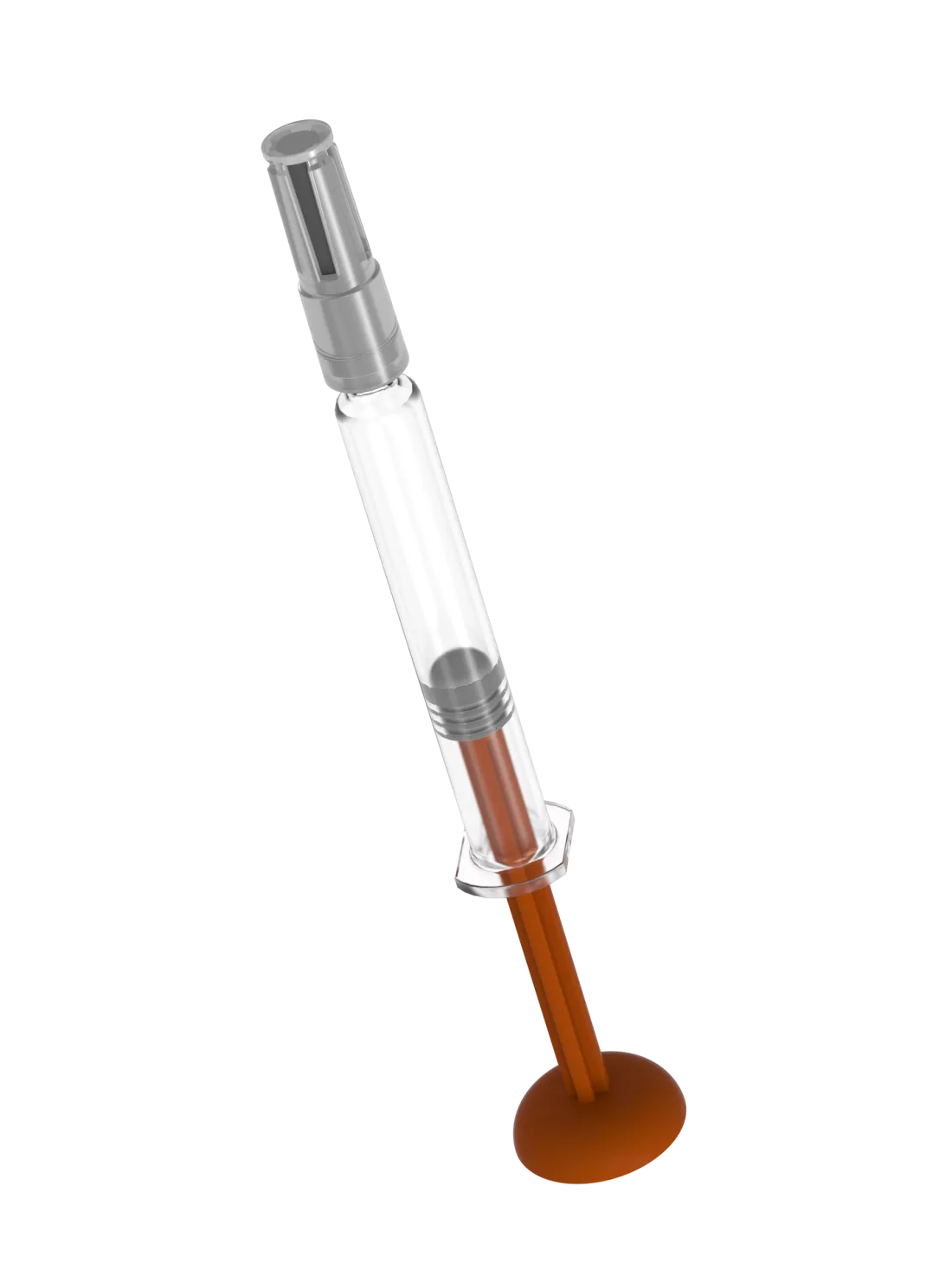
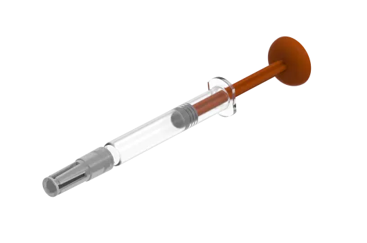
Clogged syringe needles in prefilled syringes (PFS)
Prefilled syringes (PFS) are increasingly used for medical treatment, e.g. with monoclonal antibodies. In very rare cases, the needles of prefilled syringes can become clogged. This can have detrimental consequences for patients if their medication does not enter the body or the dosage is too low. Precisely analysing and identifying the causes is crucial to solve these problems.
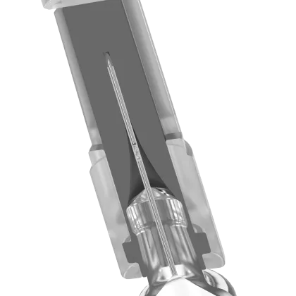
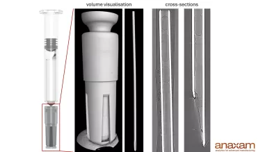
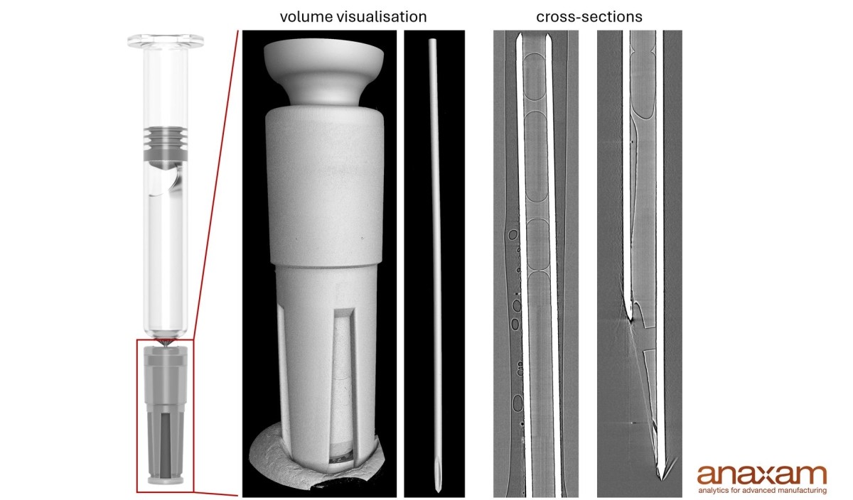
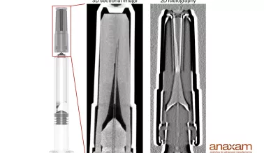
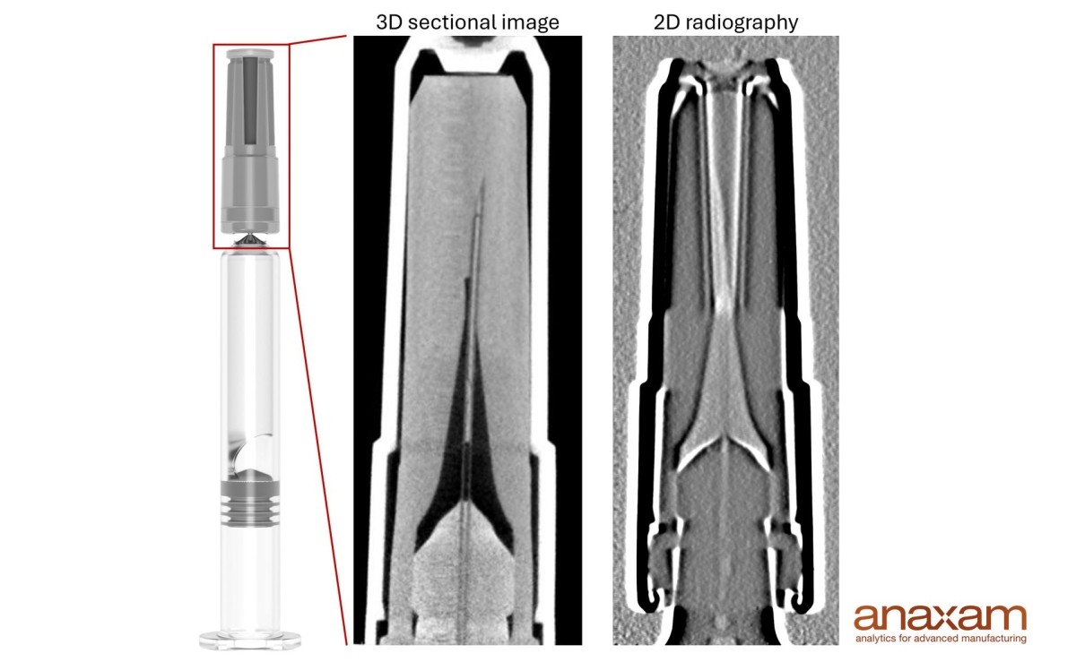
High-resolution 3D visualisation of the blockage using Synchrotron CT
Synchrotron CT is a high-resolution 3D Imaging method for analysing interfaces between air and liquid inside a needle. The method allows over 100 syringes to be analysed per day.
3D Synchrotron CT
2D visualisation of the blockage using Neutron Radiography
Neutron Radiography is a 2D Imaging method for analysing the presence of drugs inside a needle. The method makes it possible to examine over 100 syringes per day.
2D Neutron Radiography
These customers trust our expertise
Client logos are shown with prior approval.


Answers to the most frequently asked questions
Our analytical methods can be applied to all types of pre-filled syringes and autoinjectors.
Clogging is detected by high-resolution Synchrotron CT and Neutron Radiography.
Neutron Radiography enables two-dimensional visualisation of the fluid within the needle under varying environmental conditions, which helps to identify the causes of clogging.
Synchrotron CT offers high-resolution three-dimensional imaging that provides detailed insights into the interfaces between air and liquid within the needle and can thus reveal the causes of clogging.
Yes, of course. We are here to advise you without obligation and to find out whether we can support you. Please do not hesitate to contact us.
Insights from customer projects
These projects show how we work with our customers to analyze complex material challenges and generate relevant insights using neutron and synchrotron techniques.
“Visualizing how injected medications behave within tissue is a key insight for understanding the impact a clogged needle can have on the injection process. Synchrotron X-ray tomography makes this possible in a truly impressive way. The 3D images provide even more detailed information. The outstanding expertise of the entire interdisciplinary team made this achievement possible.”
Dr. Alexander Zürn, Associate Director Advanced Testing Global Device & Packaging Development,
NovartisThe combination of neutron imaging and synchrotron X-ray tomography provided valuable insights into the dynamics of liquid movement in staked-in-needle pre-filled syringes. The detailed morphological analysis enhanced the understanding of microstructural arrangements within the needle. This research contributes to addressing the issue of needle clogging and can guide the development of strategies to improve pre-filled syringe performance significantly.”
Dr. Guangli Hu, Principal Scientist, Device Development,
Merck & Co., Inc.Using the advanced methods offered by ANAXAM enables us to gain new perspectives in the field of imaging techniques. With the help of Neutron radiography and Synchrotron CT, we want to expand our knowledge of making the next generation of drug containment and delivery solutions safer and easier to use.”
Dr. Liliya Vladislavova, Product Engineer,
SCHOTT PharmaDiscuss your challenge with us
Industrial challenges can be highly specific. We’d be happy to discuss your individual case in detail. Schedule a meeting with us to learn how we can contribute to finding the right solution.

Insights from applied research

Investigating Zinc Migration from Rigid Needle Shield to Drug Formulation in Needle Tip of Pre-filled Syringe
Does needle clogging change the spatial distribution of injected drug in tissue? New insights by X-ray computed tomography
Unraveling Pre‑filled Syringe Needle Clogging: Exploring a Fresh Outlook Through Innovative Techniques
Tailor-made infrastructure for real-time investigation of prefilled syringes under varying temperature and pressure conditions using Synchrotron CT

Tailor-made infrastructure for the controlled injection of prefilled syringes (PFS) into tissue samples
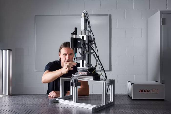
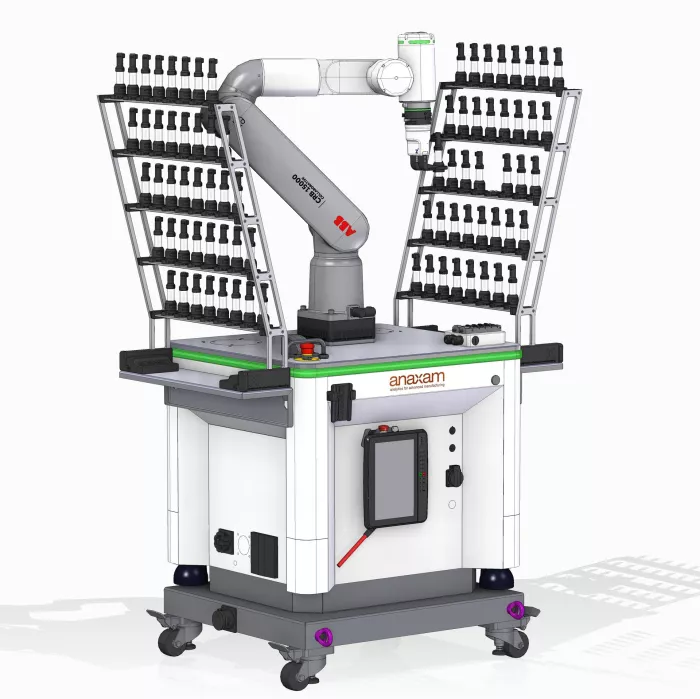
Fully automated sample changing unit using robotic infrastructure
Discover further solutions


Your access to large-scale research facilities and how we work with you
The large-scale research facilities for our material analysis


Tap the pins to explore
The way we work with you
Your
challenge
Competent
consulting
Applied material analytics with Neutron and Synchrotron radiation &
tailor-made infrastructure
Data analysis and interpretation
Final
report